Hypoechoic Nodule In Breast Meaning

Ultrasound Of Left Breast Showing A Hypoechoic Nodule With A

Ultrasound Of Hypoechoic Mass Or A Solid Breast Lump With Moose

Tubular Adenoma Of The Breast Radiological And Ultrasound Findings

An Ultrasound Of The Breast Showing A Well Defined Hypoechoic

Thyroid Nodule Sonography Assessment For Risk Of Malignancy

Breast Papillary Lesions An Analysis Of 70 Cases Ecancer

Breast Panniculitis With Vasculitis On Ultrasound A A

Breast And Axilla 5 1 Benign Lesions Case 5 1 4 Cysts Pitfalls

What Are The Breast Ultrasonography Findings Characteristic Of

Benign And Malignant Characteristics Of Breast Lesions At

Hypoechoic Nodule At The Junction Of The Left Thyroid Lobe And

Breast Mammogram Shows Mass What To Do Next

Hyperechoic Breast Lesions Radiology Reference Article

Ultrasound Images Described In Breast Papillary Lesions A

A Ultrasonography Of The Right Breast Nodule Showing Hypoechoic

Pin On Abd 300 Mod 4 Ultrasound

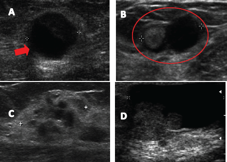
Markedly Hypoechoic Nodule Left And Hyperechoic Nodule Right

A Well Defined Hypoechoic Lesion 52 28 Mm In The Left Breast

Presentation1 Pptx Radiological Imaging Of The Thyroid Gland Disease

A On Ultrasound Imaging 1 7 2 6 Cm Irregular Illdefined
Source : pinterest.com
